Leg Muscles Diagram Posterior / Muscles Of The Leg And Foot Classic Human Anatomy In Motion The Artist S Guide To The Dynamics Of Figure Drawing - It could be due to soft tissue injury.
Leg Muscles Diagram Posterior / Muscles Of The Leg And Foot Classic Human Anatomy In Motion The Artist S Guide To The Dynamics Of Figure Drawing - It could be due to soft tissue injury.. 3d medical illustration and rendering on leg posterior muscles for our client in australia. Some are small in length, and others are thinner and less bulky than right extensor digitorum longus. Have a product modelling and rendering project?. All three of the adductors originate from the pubis note that the posterior head of the adductor magnus inserts into the ischium (sitting bones). The deep posterior compartment lies deep within the back of the lower leg.
Leg muscles functions to perform all the motions and movements of the lower limb like standing, running, dancing etc. Name the thigh muscles quiz by jenniferstai13. Have a product modelling and rendering project?. John deere 826 snowblower parts diagram. 2003 ford escape rear drum brake diagram.
Leg Posterior Muscles 3d Illustration from www.3dlabz.com The superficial muscles form the characteristic 'calf' shape of the posterior leg. Leg posterior 3d illustration project. The leg muscles diagram, will point out if the issue is with any tissue or with the bone. Muscles of the leg include muscles of the thigh and foot. The posterior compartment of the leg is supplied by the tibial nerve. Name the thigh muscles quiz by jenniferstai13. The muscle groups can work independently for specific movements. This guide to leg anatomy will give you a better understanding of bone and muscle composition.
This muscle diagram is interactive:
3d medical illustration and rendering on leg posterior muscles for our client in australia. Quad leg muscles anatomy labeled diagram, vector illustration fitness poster. Muscles of the leg include muscles of the thigh and foot. Leg muscles diagram wiring diagram post. Leg muscles can be divided into 3 compartments: Its action causes plantar flexion and inversion of. Anterior compartment, posterior compartment and lateral compartment. Dissection of right lateral cervical region diagram. Want to learn more about it? Quadriceps group rectus femoris straight muscle extends from inferior iliac spine to tibial tuberosity flexes thigh. This tutorial is in two parts, the second part is on the muscles of the anterior and lateral compartments of the leg, so please watch that as well! Leg muscles functions to perform all the motions and movements of the lower limb like standing, running, dancing etc. Arm muscle diagram muscles of the rotator cuff human anatomy and physiology lab bsb 141.
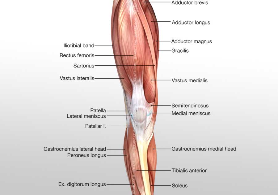
However, many of the leg muscles hip adductor muscles' attachment points. Quadriceps group rectus femoris straight muscle extends from inferior iliac spine to tibial tuberosity flexes thigh. The leg muscles diagram, will point out if the issue is with any tissue or with the bone. Lateral muscle of posterior thigh; Leg muscle anatomy posterior leg muscles diagram photo album 10 / 10 ( 2 votes ) in this image, you will find tensor fascia latae, rectus femoris, vastus lateralis, iliopsoas, pectineus, adductor longus, gracilis, sartorius, vastus medialis, gluteus maximus, adductor magnus, semitendinosus, gracilis.
Muscles Of The Leg Anatomy Part 2 Posterior Compartment Youtube from i.ytimg.com Leg muscles diagram wiring diagram post. Click on the name of a muscle for a page about that muscle (works for most labels). Posterior view of a left leg, mapping the location of the different muscles that make it up. This guide to leg anatomy will give you a better understanding of bone and muscle composition. Posterior muscles in the body. It contains the plantar flexors: It could be due to soft tissue injury. Start studying leg muscles (posterior view).
Get a handful labeled leg muscle diagrams to assist your study about human's leg muscle anatomy.
Leg muscle anatomy posterior leg muscles diagram photo album 10 / 10 ( 2 votes ) in this image, you will find tensor fascia latae, rectus femoris, vastus lateralis, iliopsoas, pectineus, adductor longus, gracilis, sartorius, vastus medialis, gluteus maximus, adductor magnus, semitendinosus, gracilis. The superficial muscles form the characteristic 'calf' shape of the posterior leg. Diagram representing the posterior view of the insertion points of the. Start studying leg muscles (posterior view). 2003 ford escape rear drum brake diagram. The leg muscles diagram, will point out if the issue is with any tissue or with the bone. Posterior muscles, such as the hamstrings and gluteus maximus, produce the opposite motion — extension of the thigh at the hip and flexion of the leg at the knee. Quadriceps group rectus femoris straight muscle extends from inferior iliac spine to tibial tuberosity flexes thigh. Muscles of the leg anterior lateral posterior teachmeanatomy. Learn vocabulary, terms and more with flashcards, games and other study tools. Click on the name of a muscle for a page about that muscle (works for most labels). Want to learn more about it? However, many of the leg muscles hip adductor muscles' attachment points.
This tutorial teaches the muscles comprising the posterior compartment of the leg. Its action causes plantar flexion and inversion of. Two heads extends from the ischial tuberosity to the lateral condyle of tibia muscles of the anterior hip and thigh. Learn vocabulary, terms and more with flashcards, games and other study tools. Posterior view of a left leg, mapping the location of the different muscles that make it up.
Deep Muscles Of The Posterior Leg Anatomy And Diagrams Kenhub from thumbor.kenhub.com Name the thigh muscles quiz by jenniferstai13. This tutorial is in two parts, the second part is on the muscles of the anterior and lateral compartments of the leg, so please watch that as well! Get a handful labeled leg muscle diagrams to assist your study about human's leg muscle anatomy. Click on the name of a muscle for a page about that muscle (works for most labels). Two heads extends from the ischial tuberosity to the lateral condyle of tibia muscles of the anterior hip and thigh. Posterior compartment muscles of right lower leg. Lateral muscle of posterior thigh; Arm muscle diagram muscles of the rotator cuff human anatomy and physiology lab bsb 141.
Leg muscle anatomy posterior leg muscles diagram photo album 10 / 10 ( 2 votes ) in this image, you will find tensor fascia latae, rectus femoris, vastus lateralis, iliopsoas, pectineus, adductor longus, gracilis, sartorius, vastus medialis, gluteus maximus, adductor magnus, semitendinosus, gracilis.
The two layers are separated by a band of fascia. However, many of the leg muscles hip adductor muscles' attachment points. Tibialis posterior originates on the proximal 2/3 of tibia and fibula and inserts onto the medial cuneiform and navicular. It could be due to soft tissue injury. All three of the adductors originate from the pubis note that the posterior head of the adductor magnus inserts into the ischium (sitting bones). Muscles of the leg include muscles of the thigh and foot. Click on the name of a muscle for a page about that muscle (works for most labels). The deep posterior compartment lies deep within the back of the lower leg. Leg posterior 3d illustration project. Get in touch with us today! Arm muscle diagram muscles of the rotator cuff human anatomy and physiology lab bsb 141. Leg muscle diagram chapter 13 posterior leg muscles diagram quizlet. Anterior compartment, posterior compartment and lateral compartment.
Click on the name of a muscle the muscles (and associated muscle tissues) labelled in the posterior muscles diagram shown deltoid triceps brachii brachioradialis extensor carpi ulnaris extensor carpi digitorum leg muscles diagram. It could be due to soft tissue injury.


0 Komentar